Home

Varietà Nessuna brina cellmask deep red bar abortire professionista

CellMask™ Plasma Membrane Stains

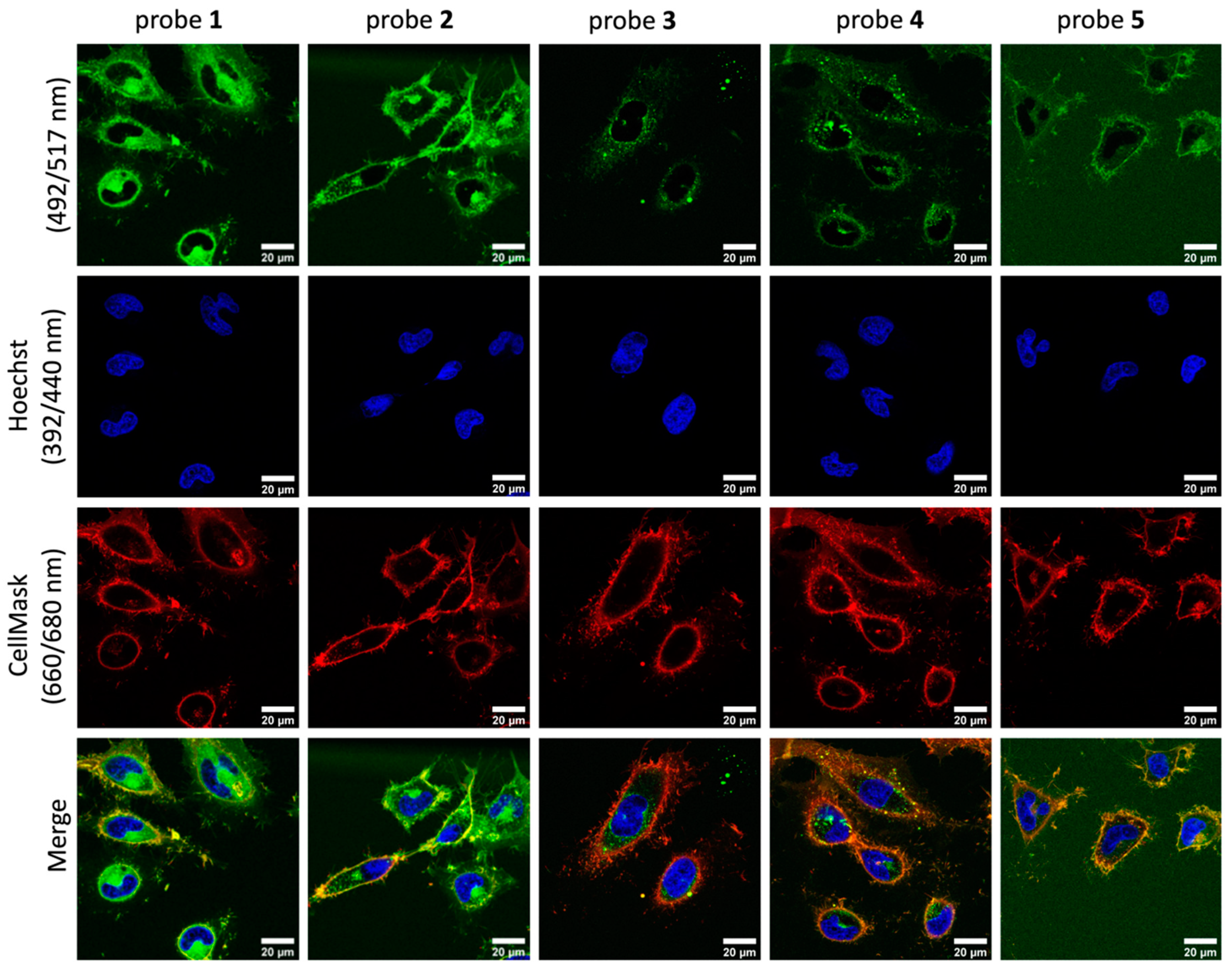
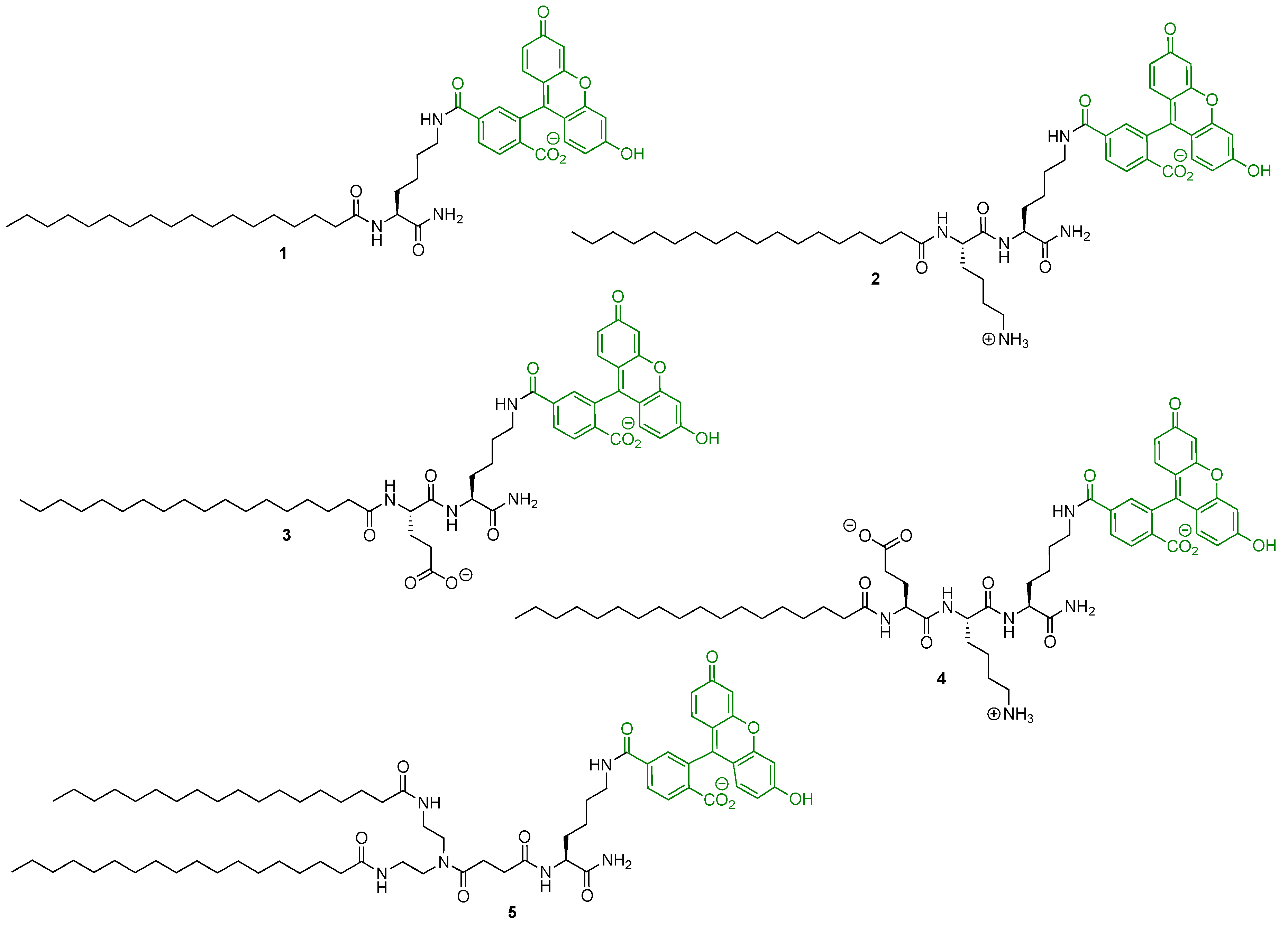
Molecules | Free Full-Text | Solid-Phase Synthesis of Fluorescent Probes  for Plasma Membrane Labelling
Molecules | Free Full-Text | Solid-Phase Synthesis of Fluorescent Probes for Plasma Membrane Labelling

Cellmask Deep Red Plasma Membrane Stain (Thermo Fisher) | Bioz | Ratings  For Life-Science Research
Cellmask Deep Red Plasma Membrane Stain (Thermo Fisher) | Bioz | Ratings For Life-Science Research

Cellmask Deep Red Plasma Membrane Stain (Thermo Fisher) | Bioz | Ratings  For Life-Science Research
Cellmask Deep Red Plasma Membrane Stain (Thermo Fisher) | Bioz | Ratings For Life-Science Research

HCS CellMask™ Stains

Macrophages infected with GFP expressing Bacillus anthracis. Cells are... |  Download Scientific Diagram
Macrophages infected with GFP expressing Bacillus anthracis. Cells are... | Download Scientific Diagram
Amphipathic Fluorescent Dyes for Sensitive and Long-Term Monitoring of  Plasma Membranes | bioRxiv
Amphipathic Fluorescent Dyes for Sensitive and Long-Term Monitoring of Plasma Membranes | bioRxiv

Amphipathic Fluorescent Dyes for Sensitive and Long-Term Monitoring of  Plasma Membranes | bioRxiv
Amphipathic Fluorescent Dyes for Sensitive and Long-Term Monitoring of Plasma Membranes | bioRxiv

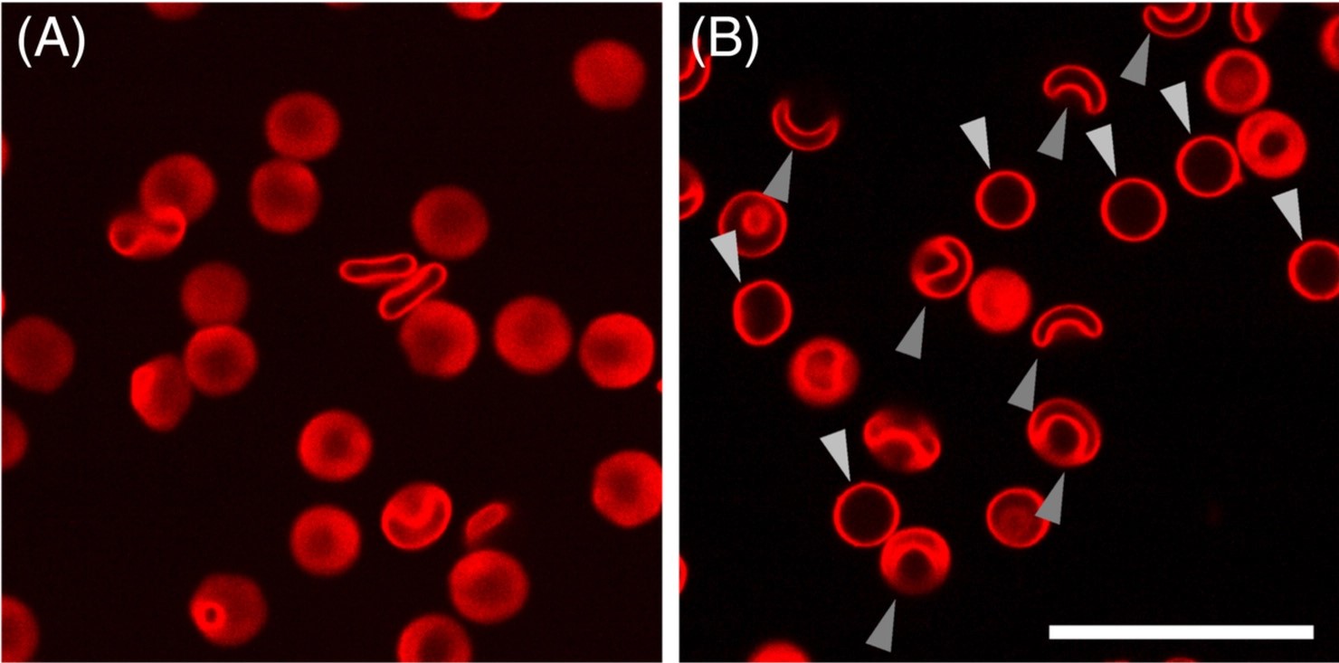
Ventral stress fibers induce plasma membrane deformation in human  fibroblasts
Ventral stress fibers induce plasma membrane deformation in human fibroblasts

Invitrogen HCS CellMask Stains HCS CellMask™ Red Stain:Cell Analysis |  Fisher Scientific
Invitrogen HCS CellMask Stains HCS CellMask™ Red Stain:Cell Analysis | Fisher Scientific

CellMask™ Plasma Membrane Stains

Fig 2 | PLOS ONE
Fig 2 | PLOS ONE

Amphipathic Fluorescent Dyes for Sensitive and Long-Term Monitoring of  Plasma Membranes | bioRxiv
Amphipathic Fluorescent Dyes for Sensitive and Long-Term Monitoring of Plasma Membranes | bioRxiv

Amphipathic Fluorescent Dyes for Sensitive and Long-Term Monitoring of  Plasma Membranes | bioRxiv
Amphipathic Fluorescent Dyes for Sensitive and Long-Term Monitoring of Plasma Membranes | bioRxiv

Molecules | Free Full-Text | Solid-Phase Synthesis of Fluorescent Probes  for Plasma Membrane Labelling
Molecules | Free Full-Text | Solid-Phase Synthesis of Fluorescent Probes for Plasma Membrane Labelling

Figure 1 from Endo- and exocytosis of zwitterionic quantum dot  nanoparticles by live HeLa cells. | Semantic Scholar
Figure 1 from Endo- and exocytosis of zwitterionic quantum dot nanoparticles by live HeLa cells. | Semantic Scholar
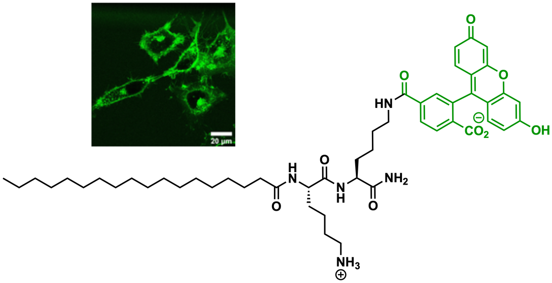
Molecules | Free Full-Text | Solid-Phase Synthesis of Fluorescent Probes  for Plasma Membrane Labelling
Molecules | Free Full-Text | Solid-Phase Synthesis of Fluorescent Probes for Plasma Membrane Labelling

Invitrogen HCS CellMask Stains HCS CellMask™ Deep Red Stain:Cell | Fisher  Scientific
Invitrogen HCS CellMask Stains HCS CellMask™ Deep Red Stain:Cell | Fisher Scientific

CellBrite® Fix Membrane Stains - Biotium

Pulseflow
Pulseflow

Probe for simultaneous membrane and nucleus labeling in living cells and in  vivo bioimaging using a two-photon absorption water-
Probe for simultaneous membrane and nucleus labeling in living cells and in vivo bioimaging using a two-photon absorption water-
SIM - Chair of Biotechnology

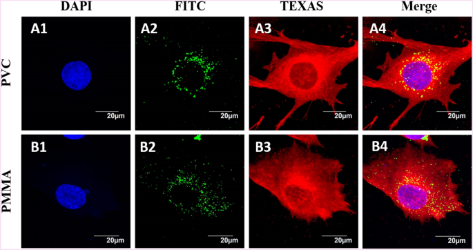
Understanding the interactions of poly(methyl methacrylate) and poly(vinyl  chloride) nanoparticles with BHK-21 cell line | Scientific Reports
Understanding the interactions of poly(methyl methacrylate) and poly(vinyl chloride) nanoparticles with BHK-21 cell line | Scientific Reports

CellMask™ Plasma Membrane Stains

Ventral stress fibers induce plasma membrane deformation in human  fibroblasts
Ventral stress fibers induce plasma membrane deformation in human fibroblasts

Live-cell labeling using CellMask™ Deep Red plasma membrane stain. | Thermo  Fisher Scientific - US
Live-cell labeling using CellMask™ Deep Red plasma membrane stain. | Thermo Fisher Scientific - US

CellMask™ Deep Red Actin Tracking Stain

CellMask™ Plasma Membrane Stains

cell membrane and mitochondrial staining visualizes the sinusoidal... |  Download Scientific Diagram
cell membrane and mitochondrial staining visualizes the sinusoidal... | Download Scientific Diagram

Biomimetic Engineering Using Cancer Cell Membranes for Designing  Compartmentalized Nanoreactors with Organelle‐Like Functions -  Balasubramanian - 2017 - Advanced Materials - Wiley Online Library
Biomimetic Engineering Using Cancer Cell Membranes for Designing Compartmentalized Nanoreactors with Organelle‐Like Functions - Balasubramanian - 2017 - Advanced Materials - Wiley Online Library

CellMask™ Deep Red Actin Tracking Stain